

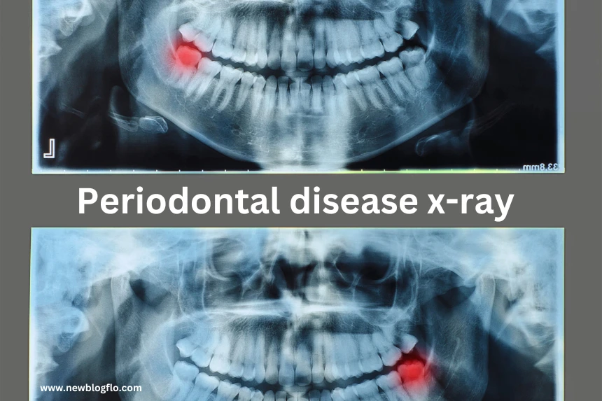
Retain your smile, protect your health, and make informed dental choices with our fail-proof guide to periodontal disease X-ray examinations. Periodontal (gum) disease is a serious condition that can lead to tooth loss if left untreated.
The early stages often show no obvious symptoms, which is why regular dental x-rays are critical for detection and monitoring. X-ray imaging allows dentists to see below the gums to evaluate bone loss, tooth damage, and infection—key indicators of periodontal disease progression.
This guide will explain how x-rays are used for periodontal diagnosis, what dentists look for on the films, and why early intervention is so important. Periodontal disease affects these deep structures, and only x-rays can uncover issues not visible during a regular dental exam. Read on to learn all about using x-rays to accurately diagnose different stages of periodontal disease.
What is Periodontal Disease?
Periodontal diseases are infections caused by plaque, a sticky film of bacteria that builds up on teeth. These include gingivitis, which affects the gums, and periodontitis, which damages the bones and tissues supporting the teeth.
Symptoms include red, swollen gums, bleeding when brushing, painful chewing, loose teeth, and chronic bad breath. While gingivitis is reversible with professional cleanings, periodontitis requires more intensive treatments to stop its progression. The earlier periodontal disease is caught, the better the outcome.
Types of Periodontal Diseases
Gingivitis: This mild and reversible form of gum disease causes red, swollen gums that may bleed easily. X-rays typically appear normal, but clinical exams reveal inflammation. Daily plaque removal and regular cleanings can usually resolve gingivitis.
Early periodontitis: Bone loss less than 15% At this stage, x-rays start to show early signs of bone loss around the teeth. There may be slight gum recession and deeper periodontal pockets. Scaling, root planing, and more diligent home care can often arrest further progression.
Moderate periodontitis: Bone loss of 15–33% Ongoing bone loss is apparent on x-rays, with pockets around teeth measuring up to 6mm. The gums recede more and may bleed easily. At this stage, surgical treatment may be needed to stop disease progression.
Severe periodontitis: Bone loss exceeding 33% but not reaching the tooth roots Involves extensive loss of connective tissue and bone support. This can lead to tooth mobility and loss if treatment is not promptly initiated.
Advanced Periodontitis: X-rays reveal severe bone loss near the tooth roots along with deep pockets exceeding 6mm. Teeth may loosen or shift. Extensive treatments like flap surgery and bone grafts are needed to try to save teeth.
It’s crucial to detect periodontal disease early while still in the gingivitis stage. If not treated promptly, it can progress to irreparable bone damage and tooth loss. X-rays are critical for identifying the stage so appropriate treatment can begin before extensive damage occurs.
X-Ray Imaging for Periodontal Disease
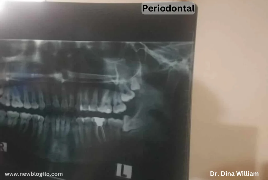
The periodontal ligament and alveolar bone surrounding teeth cannot be seen with the naked eye. Intraoral bitewing, periapical, and occlusal x-rays show bone levels, calculus, furcation involvement, and periapical lesions. Panoramic images visualize all teeth and surrounding bone in one view.
Digital radiography provides outstanding image quality for diagnostic accuracy. Using x-rays enables early detection of periodontitis before major visible damage occurs. However, they have limitations in assessing soft tissues, so a comprehensive clinical exam is also vital.
X-Ray Imaging for Detection
Dentists have several x-ray tools for identifying periodontal issues:
- Bitewing x-rays: Show bone levels between teeth and help detect bone loss indicative of periodontitis. Typically taken annually.
- Periapical x-rays: Reveal entire tooth root and surrounding bone. Used to monitor specific teeth and evaluate periodontal disease progression.
- Panoramic x-rays: Provide a broad view of all teeth, jaws, and bones in a single image. Useful for generalized screening of periodontal disease.
What Do X-Rays Reveal About Periodontal Disease?
X-rays allow dentists to see subtle changes in bone density and structure that can indicate periodontal disease:
- Widened periodontal ligament space: the healthy space between the tooth root and bone is narrowed.
- Bone loss and destruction: loss of distinct lamina dura lining around tooth sockets.
- Irregular or notched bone: Damage from inflammation and pocket formation.
- Open bone defects: Holes in bone from advanced destruction.
How Are X-Rays Used to Diagnose Periodontal Disease?
Dentists analyze x-rays for these signs of bone loss and combine this data with a visual gum examination and periodontal probing to determine a diagnosis and classify disease severity.
Key diagnostic steps include:
- Evaluating bone levels around teeth: Compare the current x-ray with previous ones to monitor bone level changes over time.
- Assessing extent of damage: Note which areas are affected and the severity of bone loss.
- Measuring pocket depths: Combine x-ray bone loss with probing measurements to gauge total damage.
- Developing a treatment plan: Match treatment options to disease severity and localization.
The Periodontal Disease X-Ray Process
Taking x-rays to aid in periodontal disease diagnosis follows a set process:
- Placement of sensor or film in mouth: Held in place by a tab while the patient bites down.
- Beam exposure at angles: X-ray tube head precisely positioned around mouth for needed views.
- Digital capture of images: Sensors communicate with computers to produce instant images.
- Protection with shielding: A lead apron and thyroid collar are used to limit radiation exposure.
- Multiple images taken: Several bitewing or periapical x-rays from different angles.
Patients can expect some pressure on biting surfaces during the procedure. Modern digital x-rays emit limited radiation and are quick to capture, making the process fast and tolerable.
Interpreting the Periodontal Disease X-Ray
Learning to correctly read an x-ray takes specialized training and an expert eye. However, here are some basics on analyzing periodontal disease x-rays:
- Compare bone levels: Look for differences between current and older x-rays for progression.
- Note angular bone defects: Narrow, sharp notches in the bone indicate active bone loss.
- Check for open margins: Separations between bone and root surfaces signal advanced disease.
- Identify furcation involvement: Bone loss around multi-rooted teeth is serious.
- Assess all surfaces: Match x-ray findings with clinical signs like pocket depths.
- Avoid overdiagnosis: Look for consistent bone patterns; bone naturally vary in density.
The Future of X-Rays in Periodontal Disease Diagnosis
While dental x-rays are integral for diagnosing periodontal disease today, emerging technologies may improve and augment their use in the future. These include:
- 3D imaging: CBCT scans provide detailed 3D views that reveal periodontal bone defects.
- Digital sensors: Continue enhancing the resolution and convenience of intraoral x-rays.
- Image processing: Software can quantify bone loss and monitor subtle changes over time.
- Laser fluorescence: Detects bacterial plaque in real-time to identify areas with the highest disease risk.
- Molecular probes: Experimental techniques to target periodontal bacteria and inflammation.
Though x-ray technology is advancing, conventional 2D dental x-rays will remain a routine part of diagnosing periodontal disease. But in the future, they will likely be supplemented by new technologies that can detect disease in its earliest phase, before bone loss even occurs.
Diagnostic Criteria
On x-rays, dentists evaluate the level of supporting bone around each tooth to determine the severity of bone loss. They also measure pocket depth by probing between the teeth and gums. This combined information indicates the stage of periodontal disease needed to plan appropriate treatment. Additional diagnostic tests, like microbiologic testing, may also be recommended by the periodontist.
Experienced dentists can identify periodontal disease indicators on x-rays before major damage occurs. Signs include:
- Widened periodontal ligament space around a tooth root
- Reduced bone height compared to previous x-rays
- Irregular or ragged alveolar bone pattern
- Bone defects between tooth roots
The amount of bone loss shown determines the stage of the disease. Dentists may use a periodontal probe to measure the exact pocket depth for confirmation. Advanced 3D scans are also being developed to provide detailed periodontal evaluations.
Treatment Options
Non-surgical treatments include deep cleanings called scaling and root planing. Antibiotics or antimicrobials may also be used. Surgical treatments like flap surgery remove tartar and infected gum tissue. Regenerative procedures can restore lost bone. X-rays are used to track treatment progress. More aggressive measures may be needed if bone loss continues.
Based on x-ray findings and clinical signs, the periodontist will determine the most appropriate treatment plan, which may include:
- Non-surgical periodontal therapy scaling and root planing
- Periodontal surgery access flap, regeneration procedures
- Antimicrobials or host response modulation
- Extraction of non-restorable teeth
- Dental implants for missing teeth
- Supportive periodontal maintenance
Preventive Measures
The key preventive measures for periodontal disease are daily plaque removal, regular professional cleanings, smoking cessation, and, most importantly, early detection through annual x-ray screenings. This allows diagnosis in the reversible early stages before major bone loss occurs.
Regular dental exams coupled with x-ray imaging are critical for finding periodontal problems so treatment can be promptly rendered. While x-rays are vital for detecting issues, prevention is ideal.
Removing plaque through brushing, flossing, and professional cleanings allows for early diagnosis and intervention through routine x-rays before major destruction happens. Periodontal disease is preventable with proper daily care and regular dental visits for exams and cleanings.
Conclusion
To avoid future tooth loss, periodontal disease must be diagnosed early. For the best treatment planning, dentists can use periodontal disease x-ray diagnosis to identify bone loss and determine the stage of periodontitis.
All adults should get yearly bitewing x-rays and complete clinical examinations to check for early symptoms of periodontal disease before irreparable harm occurs. Early treatment protects your natural teeth and smile for the rest of your life.